Modern Endoscopic Surgery allows access to the skull base through the nose, for removal of selected tumours, usually of the pituitary gland, but also other tumours or lesions of the anterior and posterior cranial fossa. Typical examples are Pituitary Adenomas, Craniopharyngiomas, selected meningiomas of the anterior skull base, and cholesterol granulomas of the petrous apex.
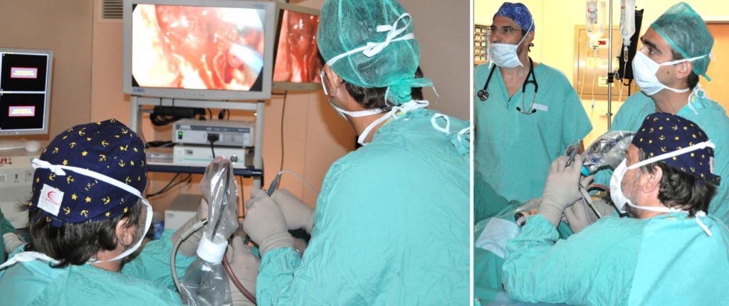
The operation is done under general anasthesia by an ENT-Neurosurgery Team. The Rhinologist and the Neurosurgeon work through the nostrils, without external incision (craniotomy). Surgical endoscopes, high resolution camera and monitors, and special microsurgical instruments are used. As in Endoscopic Surgery of the nose, the Image Guidance System (Navigator) is used, when indicated. In most cases the patient is discharged home in one or two days and is able to return to normal activities very soon.
Endoscopic surgery has dramatically reduced the morbidity of skull base operations, the complication rate and the hospitalization time, and has revolutionised modern brain surgery.
This website aims at providing simplified scientific information and not medical advice on specific conditions or individual cases. In this respect, it cannot replace the consultation and documented opinion of a specialist physician.