Skip to content Skip to footer

Hearing & Hearing Loss

How the ear works

Human ear is a little miracle of biotechnology. It has the ability to receive sound waves and convert them into electrical signals, which in turn are carried to the brain by the acoustic nerve and are presented to the higher hearing centres.

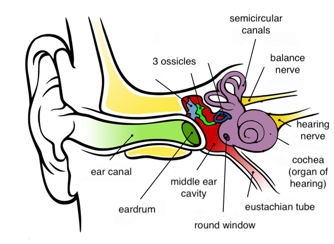
The outer ear (pinna) collects the sound waves and lead them into a slightly bent tube, the ear canal, which is obstructed, at its far end, by the elastic tympanic membrane (ear drum). Vibration of the tympanic membrane induced by the sound wave is transferred to a chain of three little bones (ossicles). The stapes, which is the last ossicle in the chain, transmits the vibration to the fluids of the inner ear, which is the main organ of hearing. Inside the inner ear, the minute hairs of the hearing cells move passively, pushed by the vibration wave. This passive movement is transduced into electrical signal inside the sensory cells. The branches of the hearing nerve collect these signals from the different hearing cells and forward them to the brain. There, after appropriate processing by the hearing centres, these signals are recognized as sound.

The space behing the ear drum (middle ear space), which hosts the three ossicles is ventilates through the postnasal space (nasopharynx) by means of a cartilaginous and osseous tube, known as the eustachian tube. Dysfunction of this tube can lead to chronic middle ear problems.


Examination of the ear under the microscope. Different magnification options allow for accurate assessment of various conditions, but also the safe performance of microsurgical office procedures, such as myringotomy (incision of the ear drum), placement of ventilation tubes, or intratympanic injections.
Normal tympanic membrane under the microscope.

How we examine the ear

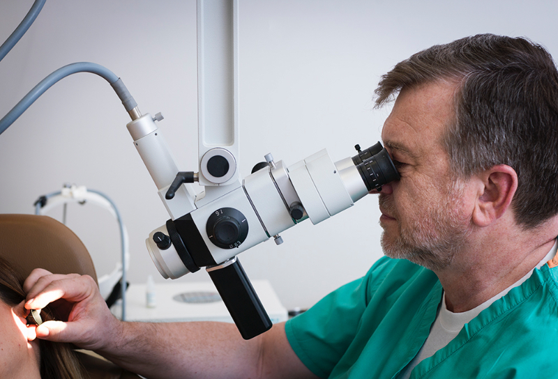
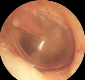
At our office, examination of the ear is always done under microscopic control. The ceiling- mounted examination microscope provides the necessary magnification for accurate diagnosis and safe micro-procedures. Ear cleaning, after certain surgeries, require fine manipulations. Minor surgical procedures, such as myringotomy, ventilation tube placement, intratympanic injections, can be performed at an office setting under local anaesthesia, saving time and hassle.

Examination of Hearing

Assessment of hearing in adults and children after 4 or 5, is done with the Pure Tone Audiogram.

Pure Tone Audiogram: Sounds of different frequencies and gradually decreasing intensities are presented to the patient, through headphones and bone conductors. The minimum audible intensity at each frequency is identified and noted on the diagram. The test is done separately for each ear.
Examples of Pure Tone Audiogram. Red curves are for right ear, blue for the left. Above: Normal test, with high position of both curves. Below: Audiogram of a 82-year-old patient, suffering from presbycusis, i.e. hearing loss due to age. Curves for both ears show a down-sloping pattern, with worse hearing in high frequencies, common finding in microvascular lesions in the organ of hearing.

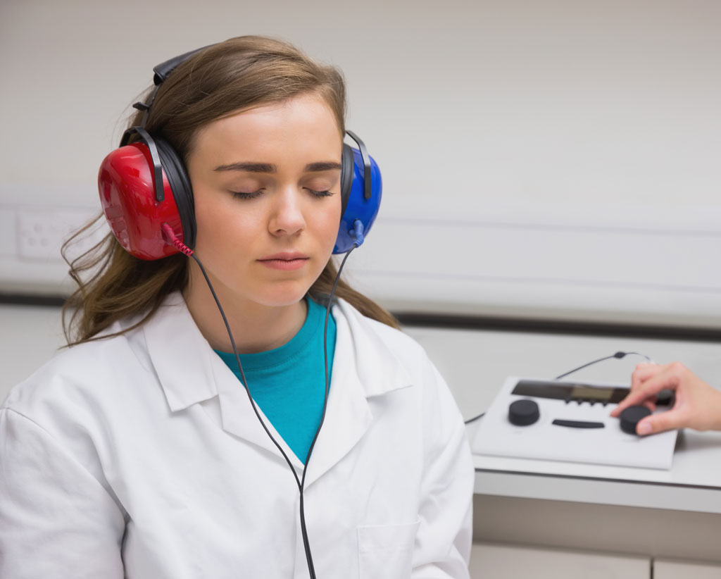
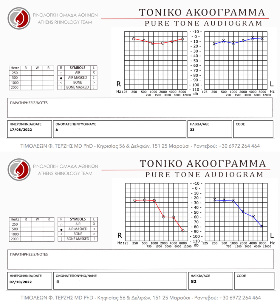
The Pure Tone Audiogram is a test which is dependent upon the replies of the examined subject. With the help of a special device, we present sound (tone) of various frequencies at decreasing intensity, and we note the lowest intensity audible at each frequency (musical note). The sound is first presented with headphones and then with a special bone conductor, which is a minute speaker, fixed on the bone behind the ear. The higher position of the curve on the diagram, the better the hearing.

Speech Audiogram. The marks on the diagram represent the percentage of words in each intensity, that can be repeated correctly by the patient. The red curve is for the right ear, the blue for the left.

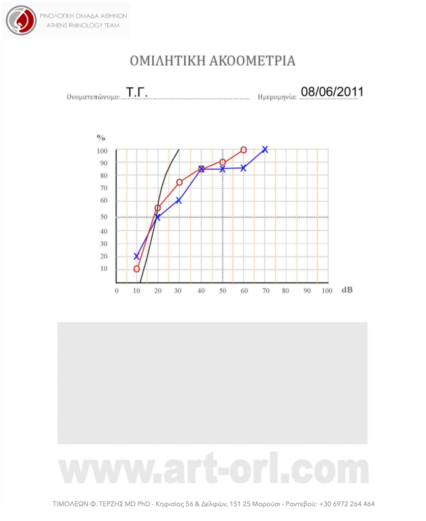
At Speech Audiogram, the examiner does not present tones to the patient, but spoken words, usually previously recorded on a CD. The intensity is gradually lowered, and the patient is asked to repeat the words he/she hears. At the Speech diagram, we note the percetage of the correctly reproduced words at each intensity level. Speech Audiogram reflects speech intelligibility, regardless of the tone audibility. It is important in specific conditions of the ear.

What can be wrong if we do not hear well

Hearing loss (or deafness) is divided into two major categories:

1. Conductive Hearing Loss
In this type of hearing loss there is a defect or dysfunction in the system which conducts sound from the environment to the inner ear, i.e. the ear canal, tympanic membrane and the three ossicles. Typical diseases which cause conductive hearing loss are wax plug in the ear canal, acute ear infection, secretory otitis media (glue ear), chronic otitis media, perforation of the tympanic membrane, cholesteatoma and otosclerosis.

A most important thing about conductive hearing loss is that, in most cases, it is not permanent, as in most cases it can be treated, either by medical or surgical means, with excellent expected results.

2. Sensori-neural Hearing Loss
This type of hearing loss can occur when there is a lesion either in the organ of hearing (inner ear) or the nerve paths involved in hearing. Characteristic examples in this category are most cases of congenital hearing loss, acoustic trauma and noise-induced hearing loss, age related hearing loss (presbycusis), hearing loss induced by drugs or infectious diseases, and sudden hearing loss.

With the exemption of sudden hearing loss, sensori-neural deafness is permanent and cannot be treated, either medically or surgically. Supportive measures, such as hearing aids or cochlear implants, may be helpful in selected cases.

In some cases, we may have mixed hearing loss, which is a combination of the two main categories mentioned above.

This website aims at providing simplified scientific information and not medical advice on specific conditions or individual cases. In this respect, it cannot replace the consultation and documented opinion of a specialist physician.

Our site uses cookies. Learn more about our use of cookies: cookie policy