How the ear works
Human ear is a little miracle of biotechnology. It has the ability to receive sound waves and convert them into electrical signals, which in turn are carried to the brain by the acoustic nerve and are presented to the higher hearing centres.
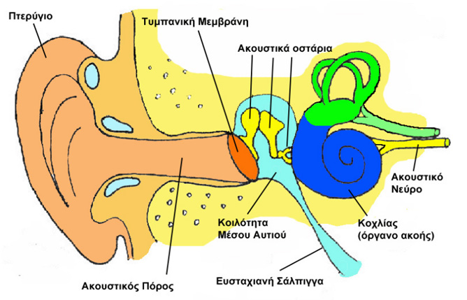
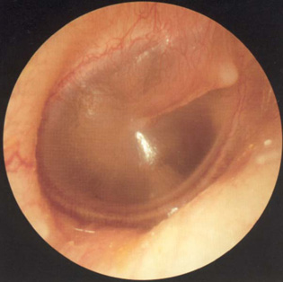
The pinna collects the sound waves and lead them into a slightly bent tube, the ear canal, which is obstructed, at its far end, by the elastic tympanic membrane (ear drum). Vibration of the tympanic membrane induced by the sound wave is transferred to a chain of three little bones (ossicles). The stapes, which is the last ossicle in the chain, transmits the vibration to the fluids of the inner ear, which is the main organ of hearing. Inside the inner ear, the minute hairs of the hearing cells move passively by the vibration wave. This passive movement is transduced into electrical signal inside the sensory cell. The branches of the hearing nerve collect these signals from the different hearing cells and forward them to the brain. There, after appropriate processing by the hearing centres, these signals are recognized as sound.
The space behing the ear drum (middle ear space), which hosts the three ossicles is ventilates through the postnasal space (nasopharynx) by means of a cartilaginous tube, known as the eustachian tube. Dysfunction of this tube can lead to chronic middle ear problems.
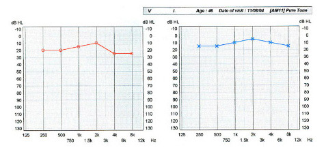
Hearing in humans can be measured with pure tone audiogram. At this subjective test, sound tones of various frequencies are presented to the patient and the lowest audible intensity at each frequency is noted on a special diagram. A separate diagram is usually drawn for each ear.
What can be wrong if we do not hear well
Hearing loss (or deafness) is divided into two major categories:
1. Conductive Hearing Loss
In this type of hearing loss there is a defect or dysfunction in the system which conducts sound from the environment to the inner ear, i.e. the ear canal, tympanic membrane and the three ossicles. Typical diseases which cause conductive hearing loss are wax plug in the ear canal, acute ear infection, secretory otitis media (glue ear), chronic otitis media, perforation of the tympanic membrane, cholesteatoma and otosclerosis.
A most important thing about conductive hearing loss is that, in most cases, it is not permanent, since it can be treated, either by medical or surgical means, with excellent expected results.
2. Sensori-neural Hearing Loss
This type of hearing loss can occur when there is a lesion either in the organ of hearing (inner ear) or the nerve paths involved in hearing. Characteristic examples in this category are most cases of congenital hearing loss, acoustic trauma and noise-induced hearing loss, age related hearing loss (presbycusis), hearing loss induced by drugs and infectious diseases, and sudden hearing loss.
With the exemption of sudden hearing loss, sensori-neural deafness is permanent and cannot be treated, either medically or surgically. Supportive measures, such as hearing aids or cochlear implants, may be helpful in selected cases.